 Current : Home page → Products
→ X-ray photoelectron spectroscopy (XPS)
Current : Home page → Products
→ X-ray photoelectron spectroscopy (XPS)
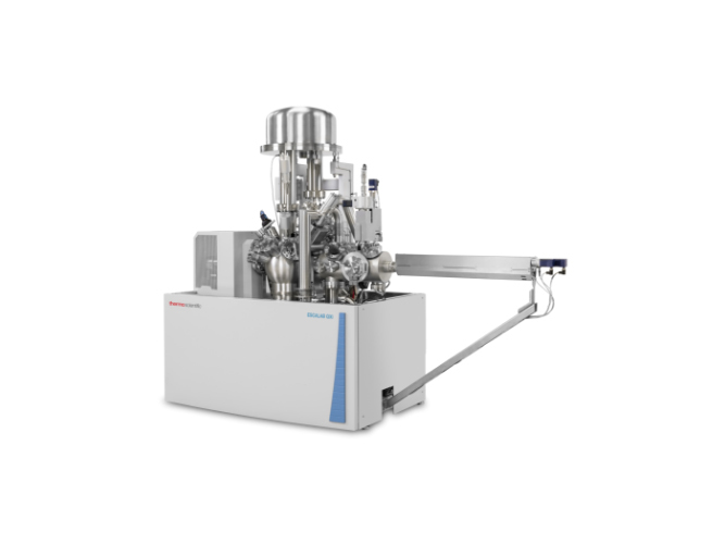


Thermo Scientific, which combines high sensitivity, high-resolution quantitative imaging, and the ability to combine multiple technologies ™ ESCALAB ™ The QXi X-ray photoelectron spectroscopy (XPS) microprobe will meet your needs for improving analytical performance and flexibility.
The ESCALAB QXi XPS microprobe is a scalable and optimized multi technology instrument with unparalleled flexibility and configurability. It is very sensitive and can produce high-quality spectra within a few seconds. System control, data collection, processing, and reporting are seamlessly integrated into the powerful Thermo Scientific Avantage data system. The cutting-edge technology driven by intuitive software ensures world-class results and productivity. The ESCALAB QXi XPS microprobe features a unique dual detector system and provides high-quality XPS imaging with excellent spatial resolution.

Thermo Scientific ESCALAB QXi Microprobe Automatic Injection
The dual crystal micro focusing monochromator comes with a 500mm Roland circle, using aluminum anode (or aluminum silver dual anode with dual monochromator option), and the test spot size can be selected in the range of 200 µ m to 900 µ m.
The electron gun coaxial with the input lens of the analyzer is used for charge compensation when analyzing non-conductive samples using a monochromatic X-ray source, while without using a magnetic lens, the dual beam electron gun generates two types of low-energy ions to assist in providing effective charge compensation and low-energy electrons.
The lens and analyzer system on ESCALAB QXi XPS microprobe has been optimized for spectroscopy and parallel imaging; A single analyzer path means that the same instrument parameters (such as energy) can be used for spectra and imaging.
The ESCALAB QXi XPS microprobe is equipped with two detector systems: one optimized for spectrum, consisting of a set of six channel electron multipliers, and the other used for parallel imaging, consisting of a pair of channel plates and a continuous position sensitive detector.
The ESCALAB QXi XPS microprobe has two options for fast and high-resolution depth analysis: the standard EX06 ion gun, optimized for single particle mode ion sputtering and ion scattering spectra; And optional single particle and cluster ion sources MAGCIS, which can be applied to single particle ion profiling, ion cluster profiling, and ion scattering spectroscopy analysis.
All analysis functions are controlled by the Avantage data system based on Windows software, which means the entire analysis process can be remotely executed as needed.
A standard block containing copper, silver, and gold samples can be used to evaluate sensitivity, set the linearity of the analyzer's energy scale, calibrate the ion source, calibrate the X-ray monochromator, and determine the conversion function of the analyzer.
All motion axes on the sample stage are controlled by the Avantage data system, and high-resolution digital cameras are installed on the instrument and accurately aligned with the analysis position.
The computer-controlled 5-axis high-precision converter (HPT) can achieve accurate sample alignment for analysis. When used in conjunction with the new automatic sample loading system, it can be used to automatically replace sample racks and run preset experimental trees.
The analysis room is composed of a 5mm thick high permeability magnetic alloy to maximize magnetic shielding efficiency, and a turbo molecular pump and titanium sublimation pump are used to extract air from the analysis room, resulting in a vacuum degree better than 5 x 10⁻¹⁰Milliba.
The standard pre-treatment room is a modular sample entry lock and preparation room with ports that can accommodate various sample preparation equipment, such as heating/cooling probes, ion guns, high-pressure gas chambers, sample parking devices, and gas inlets.
The measurement coordinates can be imported from a dedicated microscope system into the Avantage data system using Thermo Scientific Maps software, enabling faster identification of the measurement area. XPS spectroscopy and imaging data can be added to Maps software for direct comparison of surface chemistry and structural information.